The relatively low cross-section of Raman scattering prevents routine application of Raman spectroscopy in cases where need arises to characterize solid state structures with thickness in the monolayer range or biomolecular samples in the range of physiological concentrations. Apart from resonant enhancement coming from inclusion of electronic transitions, the scattering efficiency can be augmented in these cases using substrates with metallic nanostructures.
Raman enhancement is coming from amplification of the probing light in the near field, when suitably coupled to the electron plasma oscillations in the metallic nanostructures. Our focus lies in mutual tuning of the probing light and the nanostructures for optimal enhancement.
|
key available techniques:
- Resonant Raman spectroscopy (CW-RRS)
with continuously tunable wavelength of excitation
- SERS in colloids and with planar substrates
Au NP and Ag NP multistep protocols, Au-Ag core-shell NP
Au and Ag anodically oxidized and/or sparked planar substrates
- ATR-Raman
surface sensitive Raman modality
both for solid samples and in microfluidic setting
- Electrochemical Raman (EC-RS)
in microfluidic setting
- Raman tweezers (in preparation)
| 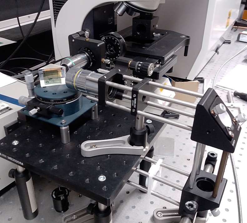 |
| equipment: | Renishaw inVia (1W @ DPPS 532nm, Streamline mapping)
Fianium WhiteLase SC450 High-Power Supercontinuum
|
|
| members: | Dusan Hemzal (SERS and Raman modalities), hemzal at physics.muni.cz
Jan Hrbac (electrochemistry), Vit Pavelka (nanoparticles, planar substrates, SERS)
|  |
| students: | Petr Steindl (FDTD), Adam Sebek (nanoparticles) |
| cooperation: | Jan Dvorak (TIRE), Karel Kubicek (biophysics)
|
| Petr Klapetek (Metrological institute; AFM, FDTD simulations), Vit Jan (Brno University of technology; SEM+EDX, AAO substrates), Anna Tycova (Inst. analytical chemistry, Czech Academy of sciences; nanoparticles), Jan Prasek (CEITEC BUT, Ag/Al2O3 planar substrates) |
|
| papers: |
Hemzal D., Prasek J., Hrdy R., Drbohlavova J., Kopp R.: SERS investigation of PO4--- to Ag/Al2O3 nanopatterned substrates binding and its dynamics for sensitive detection of phosphate in water, accepted in J. Raman Spectroscopy (2023)
Tycova A., Prikryl J., Hemzal D.: Capillary electrophoresis and Raman: can we ever expect light at the end of the tunnel?, Trac-Trends Anal. Chem. 161, 117017 (2023)
Pavelka V., Hemzal D., Hrbac J.: Complex evaluation of Raman spectra using morphological filtering: algorithms, software implementation and experimental verification of baseline correction, peak recognition and cosmic ray removal in SERS spectra of designer drugs, J. Raman Spectroscopy 53, 2100-2109 (2022)
Trachioti M.G., Hemzal D., Hrbac J., Prodromidis M.I.: Generation of graphite nanomaterials from pencil leads with the aid of a 3D positioning sparking device: Application to the voltammetric determination of nitroaromatic explosives, Sens. Actuator B-Chem. 310, 127871 (2020), link
Halouzka V., Halouzkova B., Jirovsky D., Hemzal D., et al: Copper nanowire coated carbon fibers as efficient substrates for detecting designer drugs using SERS, Talanta 165(4), 384-390 (2017), link
Sharma V., Hynek D., Trnkova L., Hemzal D., et al: Electrochemical determination of adenine using a glassy carbon electrode modified with graphene oxide and polyaniline, Microchim Acta 183(4), 1299-1306 (2016), link |
|
Older case studies:
|
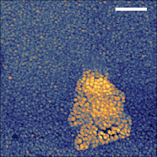
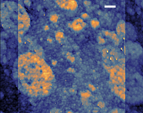
STM map of TIRE-immobilised citrate covered Ag nanoparticle(s) over PVD Au@SiO2 substrate using on-site etched Au tip; the scale bar is 5 nm. (2017)
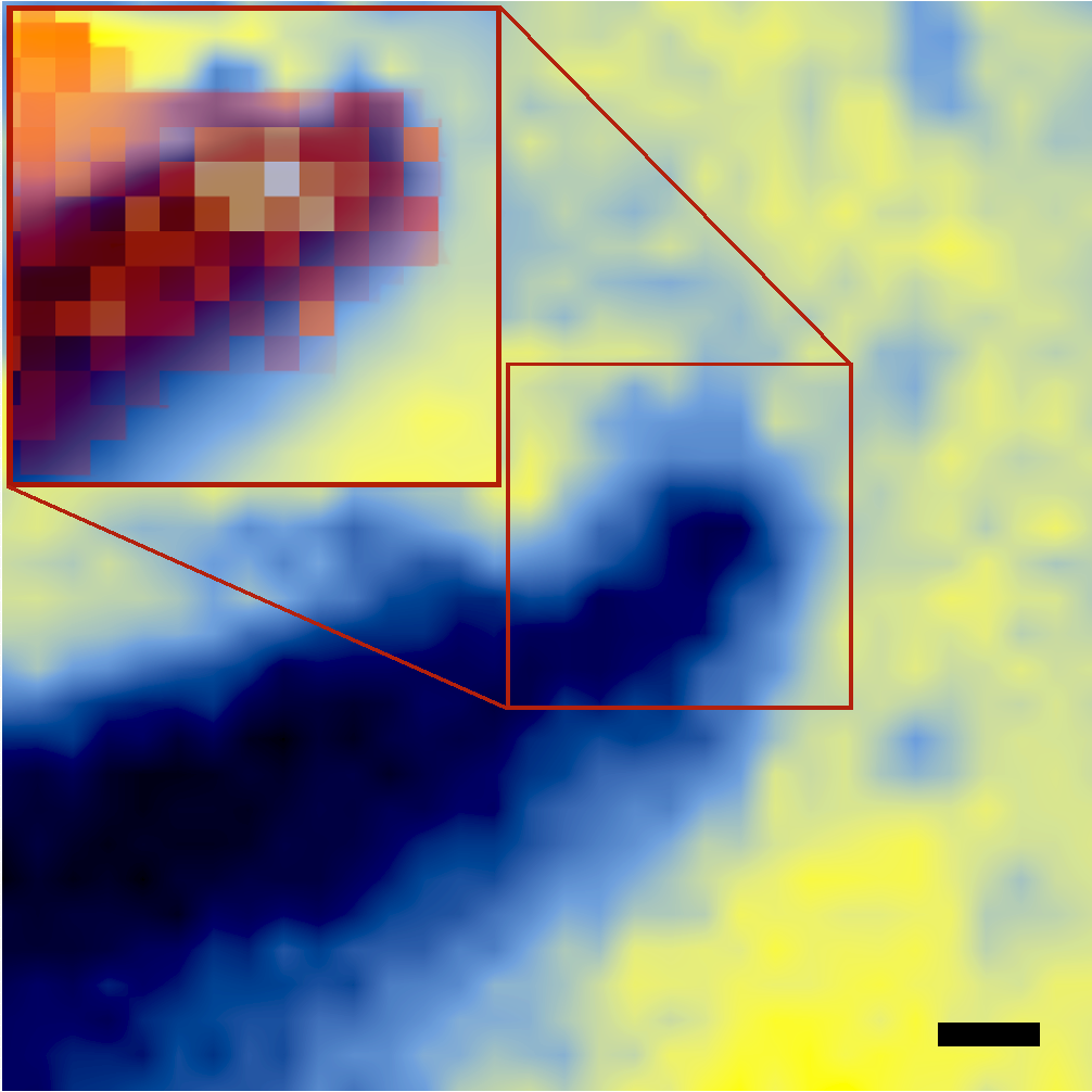
Reflectivity mapping of on-site etched Au STM tip in contact with Au@BK7 substrate at room ambient; the scale bar is 1 um. The inset shows higher resolution measurement overlayed with Raman map revealing the TERS hot spot when using 633 nm excitation. (2017)
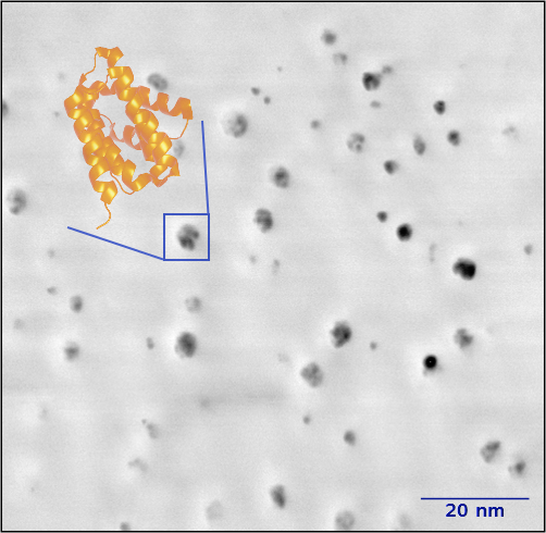
AFM phase image (air ambient) of Rtt103 protein CTD ineracting domain loaded via TIRE over Au/SiO2 substrate. Rtt103-CID is a predominantly helical peptide, some of the helices are resolved within the adsorbed molecules. The expected 2km4 structure of the peptide from pdb.org is overlayed (enlarged approx. 10x for better readability). (2016)
|
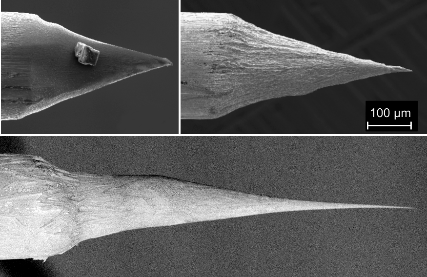
On-site etched Au STM tips with controlled angle of apex for tuning the plasmonic resonance. (2019)
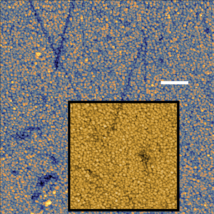
Room ambient STM map of PVD Au@BK7 substrate using on-site etched Au tip with high resolution map of a selected region included. The scale bar is 5 nm. (2017)
Dried ambient STM map of TIRE-immobilised Rtt103 protein over atomically resolved PVD Au@BK7 substrate using on-site etched Au tip; the scale bar is 1 nm. (2016)
html tabulka jako menu s klikacim ahalvim

The apex of a nose-type AFM tip before and after PVD coating with gold. (2015)
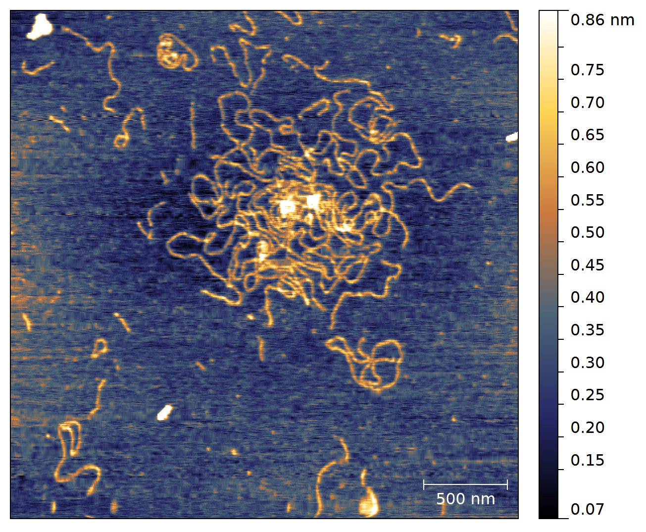
AFM scan (air ambient) of mica deposited DNA; immobilisation via Mg2+. (2014)
|
| Back to case studies |


